|
|
Rickets
- Osteomalacia during enchondral bone growth
- Age
- Histology
- Zone of preparatory calcification does not form resulting in build-up of maturing cartilage cells
- Also occurs in shafts so that osteoid production elevates periosteum
- Clinical findings
- Irritability
- Bone pain
- Tenderness
- Craniotabes
- Rachitic rosary
- Bowed legs
- Delayed dentition
- Swelling of wrists and ankles
- Location
- Metaphyses of long bones subjected to stress are particularly involved
- Imaging findings
- Cupping and fraying of metaphysis
- Poorly mineralized epiphyseal centers with delayed appearance
- Irregular widened epiphyseal plates (increased osteoid)
- Increase in distance between end of shaft and epiphyseal center
- Cortical spurs projecting at right angles to metaphysis
- Coarse trabeculation (not the ground-glass pattern found in scurvy)
- Periosteal reaction may be present
- Deformities common
- Bowing of long bones
- Molding of epiphysis
- Fractures
- Frontal bossing
- Causes Of Rickets
- Abnormality In Vitamin D Metabolism
- Associated with hyperparathyroidism
- Vitamin D deficiency
- Dietary lack of vitamin D
- Famine osteomalacia
- Lack of sunshine exposure
- Malabsorption of vitamin D
- Pancreatitis and biliary tract disease
- Steatorrhea, celiac disease, postgastrectomy
- Inflammatory bowel disease
- Defective conversion of vitamin D to 25-OH-cholecalciferol in liver
- Liver disease
- Anticonvulsant drug therapy (= induction of hepatic enzymes that accelerate degradation of biologically active vitamin D metabolites)
- Defective conversion of 25-OH-D3 to 1,25-OH-D3 in kidney
- Chronic renal failure = renal osteodystrophy
- Vitamin D-dependent rickets = autosomal recessive enzyme defect of 1-OHase
- Abnormality In Phosphate Metabolism
- Not associated with hyperparathyroidism secondary to normal serum calcium
- Phosphate deficiency
- Intestinal malabsorption of phosphates
- Ingestion of aluminum salts [Al(OH)2] forming insoluble complexes with phosphate
- Low phosphate feeding in prematurely born infants
- Severe malabsorption state
- Parenteral hyperalimentation
- Disorders of renal tubular reabsorption of phosphate
- Renal tubular acidosis (renal loss of alkali)
- deToni-Debré-Fanconi syndrome = hypophosphatemia, glucosuria, aminoaciduria
- Vitamin D-resistant rickets
- Cystinosis
- Tyrosinosis
- Lowe syndrome
- Hypophosphatemia with nonendocrine tumors
- Oncogenic rickets - elaboration of humeral substance which inhibits tubular reabsorption of phosphates
- Sclerosing hemangioma
- Hemangiopericytoma
- Ossifying mesenchymal tumor
- Nonossifying fibroma
- Hypophosphatasia
- Calcium Deficiency
- Dietary rickets = milk-free diet (extremely rare)
- Malabsorption
- Consumption of substances forming chelates with calcium
- Classification Of Rickets
- Primary vitamin D-deficiency rickets
- Gastrointestinal malabsorption
- Partial gastrectomy
- Small intestinal disease: gluten-sensitive enteropathy / regional enteritis
- Hepatobiliary disease: chronic biliary obstruction / biliary cirrhosis
- Pancreatic disease: chronic pancreatitis
- Primary hypophosphatemia; vitamin D-deficiency rickets
- Renal disease
- Chronic renal failure
- Renal tubular disorders: renal tubular acidosis
- Multiple renal defects
- Hypophosphatasia and pseudohypophosphatasia
- Fibrogenesis imperfecta osseum
- Axial osteomalacia
- Miscellaneous
- Hypoparathyroidism, hyperparathyroidism, thyrotoxicosis, osteoporosis, Paget disease, fluoride ingestion,
- ureterosigmoidostomy, neurofibromatosis, osteopetrosis, macroglobulinemia, malignancy

Rickets of the knees demonstrates
bowing of the
femurs,
metaphyseal
cupping and
fraying,
coarsening of
the trabecular
pattern,
increase in
distance
between end of
shaft and
epiphyseal
center,
poorly
ossified
epiphyseal
centers.
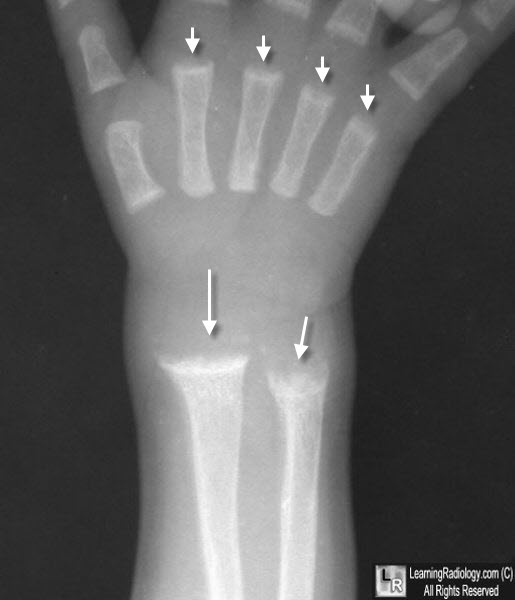
Rickets. There is cupping and fraying of all of the metaphyses (white arrows) in this skeletally-immature child.
For more information, click on the link if you see this icon 
|
|
|
