|
|
Calcinosis Universalis
-
Diffuse cutaneous, subcutaneous and
sometimes muscular calcification
-
Usually affects children and young adults
-
Not actual bone formation
-
More linear than calcifications in
scleroderma (calcinosis circumscripta)
-
Seen with dermatomyositis (polymyositis)
-
Dermatomyositis
-
Damaged chondroitin sulfate, atrophy
of muscles, followed by calcification of muscle and subcu tissue
-
Ages 5-10 and again in 50s
-
Linear and confluent calcifications
in soft tissues of extremities
-
Acro-osteolysis
-
Chest-may have infiltrates associated
-
Clinically
-
Weakness of respiratory muscles
-
Erythematous rash of eyelids
-
Proximal muscle weakness
-
Associated with a high incidence of
malignancies of GI tract, lung, ovary , breast, kidney
-
May resemble myositis ossificans
progressiva
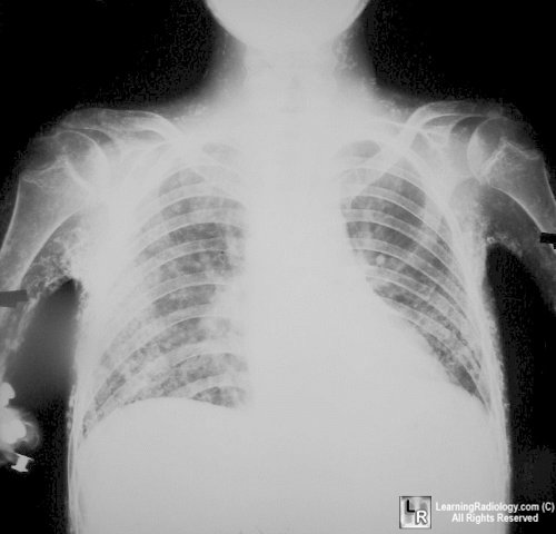
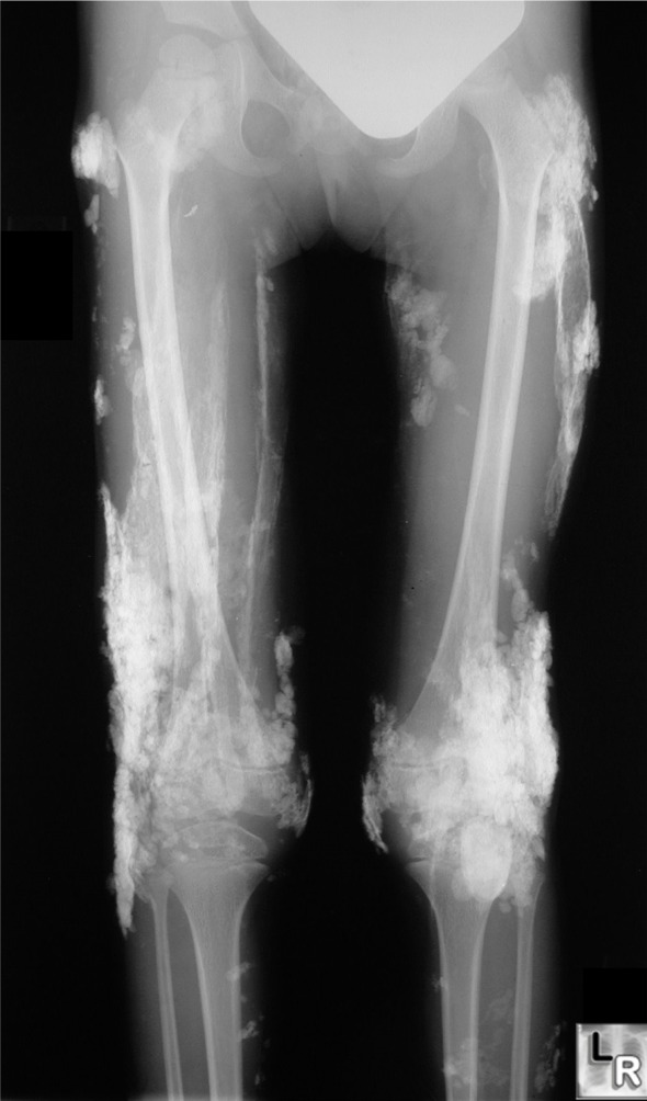
Dermatomyositis. Sheet-like calcifications seen in patients with
dermatomyositis is called calcinosis universalis because of its wide-spread distribution.
This is more likely to occur in younger patients with dermatomyositis.
For a larger photo of the same image, click on the photo above
|
|
|
