|
|
Subdural Hematoma
-
Acute hemorrhage on CT scan appears as bright on CT scan.
-
Over a period of time, the blood becomes darker.
-
Attenuation decreases by 1.5 HU per day on average.
-
There is a point when the blood becomes isodense to the brain parenchyma.
-
Contrast enhancement can help separate the clot and brain parenchyma since there is enhancement of displaced cortical vessels.
-
Subdural hematomas are usually crescentic shaped and have the capability of crossing the cranial sutures.
-
The etiology of a subdural hemorrhage occurs from a tearing of the bridging veins in the subdural space.
-
Subdural hematomas can be lethal with mortality rates ranging from 50-85%.
|
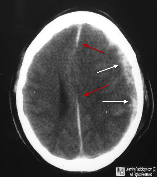
Subdural Hematoma. Axial CT scan demonstrates a left parietal subdural hematoma hyperdense to the brain (white arrows). There is associated mass effect with effacement of the left lateral ventricle and shift of the midline to the right (red arrows).
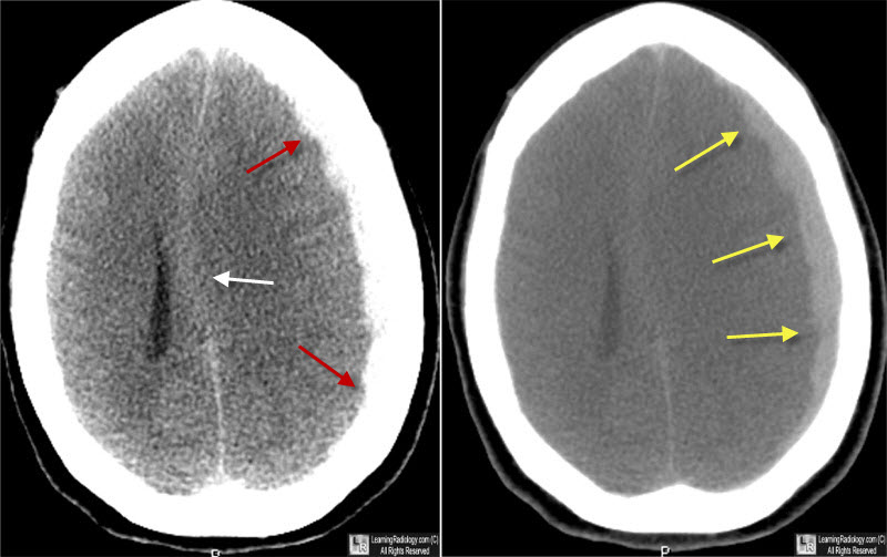
Subdural Hematoma. Image on left at brain windows shows a slight increase in density, crescentric in shape, along the inner table of the left cranial vault (red arrows) with a shift of the midline structures (white arrow) indicating mass effect. The subdural window on the right clearly shows the high density crescent-shaped blood concave toward the cerebral hemisphere (yellow arrows).
|
|
|
